Oral, Dental and Maxillofacial Radiology; also known as oral and maxillofacial radiology. It is the science of imaging methods used to diagnose diseases of the mouth, teeth, jaw bones and surrounding soft tissues.
Oral and Maxillofacial Radiology has an important place in dentistry practice because the oral structures of patients must be examined in detail in order to make a correct diagnosis.
Main Purposes of Oral, Dental and Maxillofacial Radiology:
- Detecting Dental and Jaw Diseases: Visualizing problems such as tooth decay, periodontal diseases, cysts and tumors in the jaw bone.
- Examining the Jaw Bone Structure: Diagnosis of problems such as jaw fractures, joint diseases (e.g. temporomandibular joint problems), bone loss and tooth placement disorders.
- Dental Treatment Planning: Making the correct planning required for implant placement, orthodontic treatment and other surgical interventions.
- Dental Implant and Surgery: Checking the suitability of the jaw structure before placing the implant and performing bone grafting if necessary.
- Early Diagnosis of Oral and Jaw Cancers: It is used for the early detection of jaw cancers or intraoral tumors.
Methods Used in Oral, Dental and Maxillofacial Radiology
This field uses various radiological techniques to visualize the structure of the mouth and jaw in detail. Here are some common methods:
1. X-ray
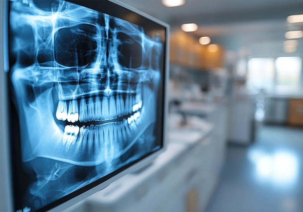
- Panoramic X-ray (Panoramic Graph): It is a common radiological examination that provides a complete image of the jaw and teeth and shows the entire mouth and jaw structure in a single film. It is especially used for tooth extraction, implant placement and orthodontic treatment planning.
- Periapical X-ray: Provides detailed visualization of the roots of the teeth, the surrounding bone structure and gum tissue. It is widely used to detect cavities, root infections or tooth root diseases.
- Bitewing X-ray: It is an x-ray technique that examines the junction areas of the upper and lower parts of the teeth. It is used to detect dental caries, especially on the contact surfaces of the upper and lower teeth.
- Jaw X-ray (Occlusal X-ray): It is an image taken to see the relationships between jaw bones and teeth. It is used in cases where tooth roots or jaw bones need to be evaluated in more detail.
2. Computed Tomography (CT or CBCT)
- Conventional Computed Tomography (CT): In addition to obtaining high-resolution cross-sectional images, it allows a more detailed examination of the jaw bones. It is used in more complex cases (for example, jaw bone diseases or large tumors).
- Digital Cone Beam Tomography (CBCT): Provides three-dimensional images of the jaw structure and is used to examine jawbone density and structure before placing dental implants. It is also very useful for orthodontic planning and jaw surgery. CBCT obtains high-resolution images using low doses of x-rays.
3. Magnetic Resonance Imaging (MRI)
- MRI is often used to evaluate soft tissues in teeth and jaw structures. It can be used for jaw joint (TMJ) diseases, tumors inside the mouth, or sinus problems. It is not preferred for imaging teeth and jaw bones because bone tissue cannot be seen clearly enough with MRI.
4. Ultrasonography
- Ultrasound may also be used occasionally for jaw joint diseases, sinus problems, or soft tissue problems. However, this method is generally not as common as radiographic (X-ray, CBCT) imaging methods.
5. Other Imaging Methods That Do Not Require Excessive Irradiation
In some cases, newer technologies such as fluorescence imaging and digital periapical devices may also be used. These methods aim to obtain clearer images with less radiation.
Application Areas of Oral, Dental and Maxillofacial Radiology
- Jaw Diseases: Detection of conditions such as tumors, cysts, infections, jaw fractures, bone development disorders and jaw joint problems in the jaw bone.
- Tooth Decay and Periodontal Diseases: It plays an important role in the diagnosis of dental caries, infections, gum diseases and periodontal disorders.
- Dental Implants: The structure, thickness and density of the jawbone are evaluated before the implant is placed. Additionally, an average layout plan is made so that the implants can be placed correctly.
- Orthodontic Evaluation: In evaluations for the correct alignment of the teeth and jaw structure, X-ray images help detect abnormalities in the jaw structure.
- Oral Cancers: X-ray or tomography is used to detect cancerous or benign tumors in the mouth in the early stages.
- Temporomandibular Joint Disorders: MRI or CT can be used to evaluate temporomandibular joint (TMJ) problems. This is important in diagnosing problems such as pain, misalignment or locking in the jaw joint.
Oral, Dental and Maxillofacial Radiology
Oral and Maxillofacial Radiology is a field where advanced imaging techniques are used to better understand many conditions in jaw structure and oral health. With methods such as x-ray, tomography and magnetic resonance, it is possible to accurately diagnose dental and jaw diseases, make correct treatment planning and provide post-treatment follow-up. Radiologists specialized in this field examine these images and make accurate diagnoses and provide guiding information to dentists or surgeons during the treatment process.

